


Euduboscquella cnemata

Diagnosis
Diagnosis_Genus: Euduboscquella Coats & Bachvaroff 2012. Euduboscquellidae with trophont episome as disc-shaped shield bordered by a perinematic ring. Lamina pharyngea extending from perinematic ring into trophont cytoplasm. Food vacuole formed as trophont emerges from host giving rise to extracellular tomont. Multiple spore morphotypes possible, including dinokont and non-dinokont cells. Individual infections producing only one type of spore.
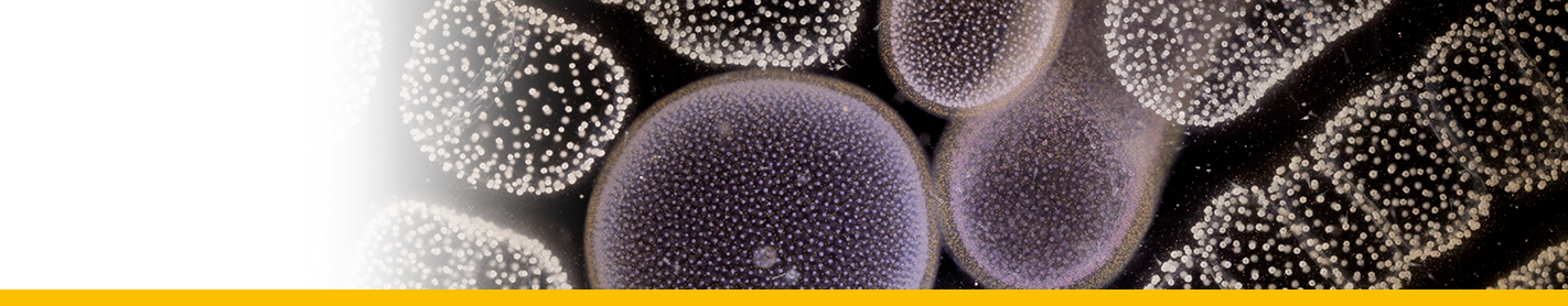
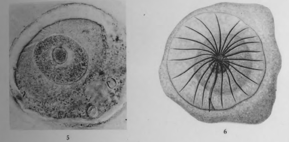
Diagnosis_Species: Trophont morphology/development: ovoid, ≤60 µm diam.; 20-30 fibers radiate from the perinema and follow furrows that spiral toward the center of the convex shield; lamina pharyngea forms a short funnel; phagotrophic stage not observed.
Sporogenesis: Occurs outside of host; sporocytes not aggregated.
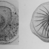
Spore morphology: One type of spore described, 3-4 µm long, similar in shape to macrospores of E. aspida.
==> E. cnemata is closed to E. aspida. It differs by the nucleolus behavior during the division and the shield appareance. The trophont is also smaller.
Body_trophont_length: ≤ 60 µm
Body_spores_length: 3 – 4 µm
Etymology
Genus name is derived from the Greek eu- (= well, normal) and the genus name Duboscquella.
Type species
The type species for the genus is E. crenulata.
Type illustration / Type locality / Type specimen
Type hosts: Favella spp.
Ecology
Substrate: endozoic (endoparasite)
Sociability_trophont: solitary
Salinity: marine
Salinity: brackish
Salinity: variable
pH: neutral
Life cycle
Live cycle of the parasite: see the development of E. aspida. The digestive vacuole (phogotrophic stage) was not observed (Cachon 1964).
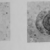
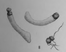
Temporary encystment of spores has been observed (Cachon 1964). They lost their flagella, and produced a hyline membrane. Rapidly, this cyst-like become turgescent, and the membrane extends as a tube. The spore is then released at one pole.
Resting_stage: cysts_asexual
Phases_alternance: haplontic
Generation: <1 month
Reproduction_mode: asexual
Symbiont: horizontal
