


Archaias angulatus
Diagnosis
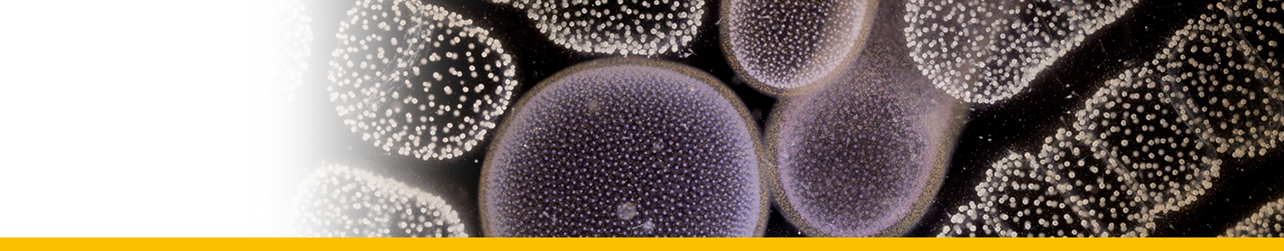
Archaias angulatus has a planispirally-coiled, flabelliform test. The overall test morphology is biconvex and lenticular in shape with a keeled periphery. The mode of coiling is initially involute with each new whorl covering previous whorls. In the terminal flabelliform stage, coiling becomes advolute (partially evolute). Archaias angulatus has a porcelaneous wall structure, in which the primary wall layer is composed of a three-dimensional random array of lath-like crystals of high-magnesium calcite. The primary wall layer is sandwiched between a thin outer veneer of flattened two-dimensionally oriented crystals that coats the surface of the chambers, and an inner basal imperforate layer from which the endoskeletal elements are formed. Each individual chamber is shaped like a coiled strip that bifurcates into two three-dimensional spirals, the alar prolongations, each of which terminates at the axis of coiling. The chambers are greater in breadth than height, and are of relatively uniform height throughout the length of the chamber. Archaias angulatus has an accretionary mode of growth, in which new chambers are formed episodically throughout the lifespan of an individual cell. Each new chamber that is formed is cemented on to the previous chamber in a separate growth event. There is minimal overlap between successive chambers; the sutures, or zones of contact between the successive chambers, are slightly depressed and spiral in shape.
The lateral surfaces of each chamber are penetrated by numerous, large, rounded pseudopores that are haphazardly distributed over most of the distal chamber face. Pseudopores, also called pits, pit-like pores or punctations , are blind pores that penetrate the shell surface, but do not perforate the test wall. The pseudopores are approximately 5-6 µm in diameter, and occur at a density of less than 1 pit per 100 µm2 of chamber surface area . An imperforate band is present along the base (proximal edge) of the chamber face adjacent to the suture.
Each chamber possesses multiple apertures that are arranged in offset rows parallel to the long axis of the apertural face . Two rows of apertures are present on each of the alar prolongations; these two sets of apertural rows merge into four or more rows of apertures on the primary apertural face . The apertures are circular, subcircular to oval in shape and are bordered by a rounded, elevated, imperforate rim. Oval-shaped apertures are oblong, with the long axis of the aperture parallel to the long axis of the apertural face. The apertural face is ornamented with a network of reticulate ridges and depressions that impart a spongy texture to the apertural face.
Endoskeletal elements present in the interior of each chamber include rounded pillars and socculi. Pillars are cylindrical or columnar structures that extend from the chamber floor to the chamber roof. Pillars (interseptal pillars or pilintradermal plates) are free-standing structures within the chamber lumen that are not connected laterally to the chamber walls. There are three-to-four rows of pillars present in terminal chambers of adult specimens of Archaias angulatus. The pillars arise from low raised ridges (socculi) that form on the floor of the chamber lumen from the basal (inner wall) layer of calcite that is deposited over the apertural face of the previous chamber. Socculi may occur as either parallel ridges or an interconnected network of ridges. The apertures of the previous chamber (intercameral foramen) are located in depressions (sulci) between the socculi. Pillars occur at regular intervals within the chamber lumen, spaced at 50-µm intervals, and are visible in live specimens or wetted dead tests viewed in transmitted light
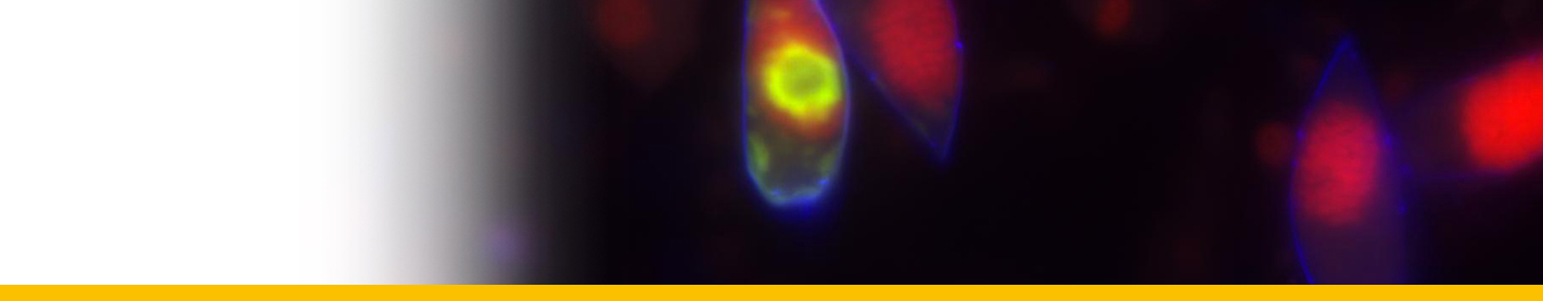
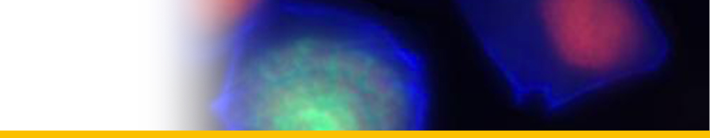
Living individuals of Archaias angulatus possess a “grass-green” coloration due to the presence of unicellular chlorophyte endosymbionts in their cytoplasm.
Reference(s)
Observation site(s)
SYMBIONTS
| Association with... | Region origin | Name of site | In reference... |
|---|---|---|---|
| Chlamydomonas hedleyi | Florida | Key Largo |
(1974) The taxonomic identity and physiological ecology of Chlamydomonas hedleyi sp. nov. algal flagellate symbiont from the foraminifer Archaias angulatus. British Phycological Journal 9:407 - 422. doi: 10.1080/00071617400650481 |
