


Dissodinium pseudolunula
Diagnosis
Diagnosis_Genus: Dissodinium Klebs in Pascher, emend. Elbr. & Dreb. 1978. Marine ectoparasitic dinophytes, host: crustacean eggs. Asexual reproduction (sporogenesis) by obligate sequence of primary cysts, secondary cysts, and formation of planospores (dinospores). Primary cyst spherical, with large food vacuole. Nucleus spherical, located in the parietal cytoplasm. Chromatophores present or lacking. Inside the primary cyst wall the protoplast divides by repeated binary fission (palintomy) into 2, 4, 8, 16, or more secondary cysts. The secondary cysts form 5 to 16 or more (rarely less than 5) dinospores. Dinospores small, Gymnodinium- or Gyrodinium-shaped, biflagellate, athecate. The dinospores attach to the host, loose their motility and form a sucker organelle. Hypertrophic growth by phagotrophic uptake of host material. Finally, the growth phase terminates with formation of a primary cyst. No bioluminescence.
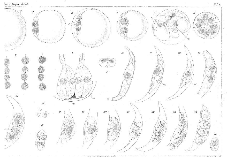
Diagnosis_Species: Dissodinium pseudolunula Swift ex Elbr. & Dreb. 1978. Ectoparasite of copepod eggs. Sporogenesis by two successive cyst stages terminating with formation of dinospores. Primary cyst spherical, 60-100-
130 µm in diameter. Cytoplasm parietal, surrounding a large vacuole filled with nutritional material. Nucleus spherical, lying in a peripheral cytoplasmic aggregation. A few greenish yellow chromatophores present, multiplying during sporogenesis. The primary cyst divides to form 8 or 16 (rarely 2 or 4) lunate secondary cysts which are liberated from the decaying primary cyst wall. The secondary cysts, about 100-140 µm from tip to tip across, contain a large nutritional vacuole. The secondary cysts form usually 5-8 (range 2-16) dinospores. Dinospores measuring 23-28 µm in length, 18-21 µm in width, athecate, biflagellate, of the Gymnodinium type, greenish yellow pigmented by numerous chromatophores, containing chl a. In each secondary cyst one dinospore contains the residue of the large vacuole as a red globule. After release from the secondary cysts the dinospores may either infest a new host or may become non-motile with following encystment. Distribution: marine plankton. When the host is absent, the species survives with a distinctive pair of twin resting cysts (previously described as Pyrocystis margalefii). The twin resting cysts are formed inside a lunate secondary sporangium, after duplication (Gomez 2013). Each infected egg may provide 32 new infective dinospores for the following spring.
Body_trophonts_length: 60-140 µm
Body_spores_length: 23-28 µm
Body_spores_wide: 18-21 µm
Body_cysts_length: 100-131 µm (Drebes 1981)
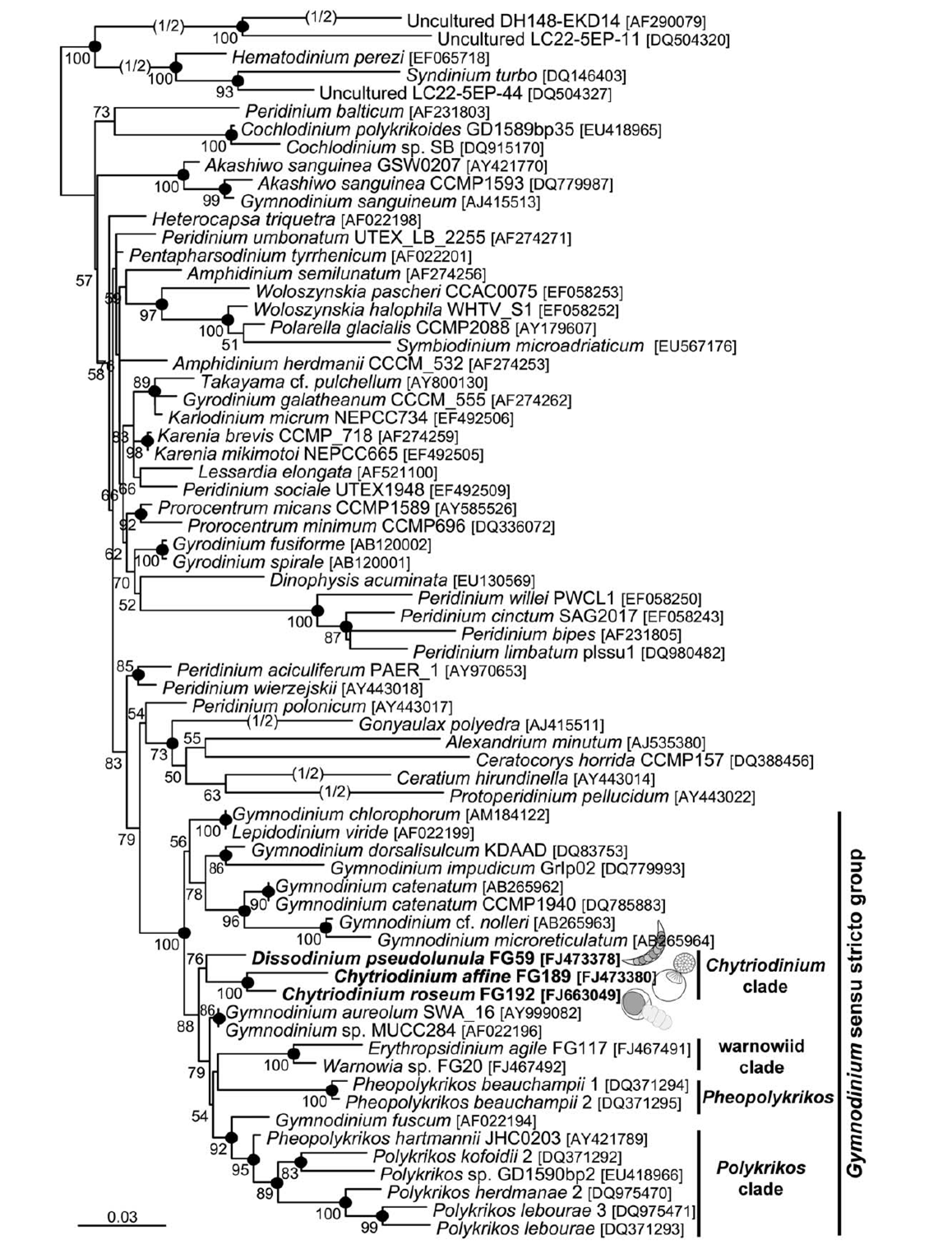
Sequence_SSU: FJ473378, FJ473379
Type species
This is the type species of the genus.
Type illustration / Type locality / Type specimen
L e c t o t y p e : Apstein, C. (1906). Pyrocystis lunula und ihre Fortpflanzung. Wiss. Meeresunters. (Abt. Kiel) 9, 263-269, P1.10 Figs lb-27.
Ecology
Substrate: epizoic
Salinity: marine
Life cycle
Generation: <1 month
Reproduction_mode: asexual
Resting_stage: cysts_asexual
Symbiont: horizontal
Feeding behaviour
Mode of locomotion
Reference(s)
Attached phylogeny
Observation site(s)
HOSTS
| Association with... | Region origin | Name of site | In reference... |
|---|---|---|---|
| Copepoda | Wimereux SOMLIT |
(2013) The formation of the twin resting cysts in the dinoflagellate Dissodinium pseudolunula, a parasite of copepod eggs. Journal of Plankton Research 35:1167 - 1171. doi: 10.1093/plankt/fbt066 |
|
| Copepoda | Marseille |
(2009) Life cycle and molecular phylogeny of the dinoflagellates Chytriodinium and Dissodinium, ectoparasites of copepod eggs. European Journal of Protistology 45:260 - 270. doi: 10.1016/j.ejop.2009.05.004 |
|
| Temora longicornis | Wadden Sea | near List/Sylt |
(1978) Dissodinium pseudolunula (Dinophyta), a parasite on copepod eggs. British Phycological Journal (European Journal of Phycology) 13:319-327. |
